 Vascular Studies
 are non-invasive
tests used to evaluate for the presence and severity of peripheral vascular disease.
BCS Heart offers on-site vascular
study evaluations including performance of ankle/brachial indices and duplex ultrasound of the aorta, carotid, subclavian,
and lower extermity arteries.
Peripheral vascular disease results from the build up of plaques within the inner walls of blood vessels. Plaques
form as a result of atherosclerosis, an inflammatory process that eventually results in calcium
deposits within blood vessel walls. If these plaques become severe enough, impairment in blood flow results.
A similar process can result in severe weakening of blood vessel walls, leading to dilated blood vessels which may
have an increased risk for rupture.
For more detailed information about vascular studies, continue
reading below.

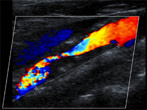
Vascular studies are usually performed by a trained technician using a combination of duplex ultrasound (see image on the
top right) and blood pressure
cuffs.
Duplex ultrasound uses sound waves emitted from a small probe on the skin surface to visualize major arteries in
the body and the blood flow within them. Very high flow velocities of blood within the artery may suggest significant
blockages that often require further evaluation with a catheterization procedure (see
Peripheral Stenting to learn more).
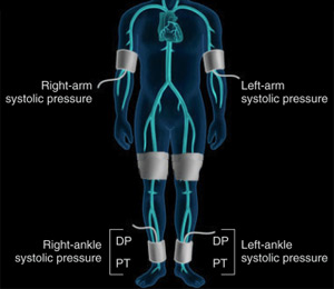
Ankle/Brachial indices are performed by measuring systolic blood pressures at
various locations of the upper and lower extremities (see image on the bottom right). This test helps to assess for hemodynamically singificant
blockages in peripheral arteries that may also require additional evaluation.

 Vascular Studies are non-invasive
tests used to evaluate for the presence and severity of peripheral vascular disease.
BCS Heart offers on-site vascular
study evaluations including performance of ankle/brachial indices and duplex ultrasound of the aorta, carotid, subclavian,
and lower extermity arteries.
 Vascular studies are usually performed by a trained technician using a combination of duplex ultrasound (see image on the
top right) and blood pressure
cuffs.