 Cardiac PET Scanning
 is a non-invasive
imaging procedure used to evaluate for abnormalities in coronary artery blood flow.
BCS Heart is one of the few
cardiology groups in central Texas to have on-site Cardiac PET scanning capabilities and to use this state-of-the-art
technology for patient care.
Red blood cells traveling through coronary
arteries are responsible for delivering oxygen to the heart in order for the heart to function appropriately.
Abnormalities, or blockages, in these arteries may exist due to coronary artery disease and impair necessary oxygen
from reaching the heart.
Cardiac PET (positron emission tomography) is a nuclear medicine imaging technique in which
pictures of the heart are obtained following the injection of a radioisotope. This test is used to assist physicians in
the diagnosis and management of coronary artery disease. PET imaging is similar to Nuclear (SPECT) Stress Testing in that both imaging techniques
involve the use of a radioisotope and the detection of gamma radiation. With PET imaging, however, the measured gamma rays
are generated from positron emission, whereas with SPECT imaging, the measured
gamma rays are directly emitted from radioisotopes. One imaging modality is not necessarily better than the other,
although higher resolution images are typically able to be generated with PET scanning.
For more detailed information about the procedure, continue
reading below.
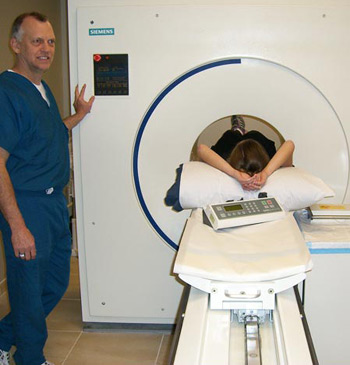
Following placement of an IV and injection of a radioisotope, images of the heart are obtained by using a specialized
scanner (see picture above). Scanning the heart takes less than 10 minutes. Following the initial scan, the patient
is then administered a medication (regadenoson) to dilate, or expand, the heart arteries to maximize blood flow to
the heart.
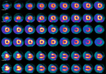 After this medication is given, a second scan of the heart is performed. The images obtained from the first
and second scans (see image to the right) are then compared and interpreted by your physician. Images are
able to provide detailed information about the function of the heart (ejection fraction), movement of the heart (wall
motion), and blood flow to the heart.
After this medication is given, a second scan of the heart is performed. The images obtained from the first
and second scans (see image to the right) are then compared and interpreted by your physician. Images are
able to provide detailed information about the function of the heart (ejection fraction), movement of the heart (wall
motion), and blood flow to the heart.
The patient is monitored by electrocardiogram throughout the procedure and the
entire procedure is completed within one hour.
Results of the procedure are typically available within 7-10 business days.